Instruments Heading link
Instrumentation and services are available to all researchers inside and outside of the university.
Researchers from Northwestern University and the University of Chicago are provided internal rates as all UIC users. Researchers from Rush University Medical Center are provided special rates as well. All other users can access services but at an external academic or external non-academic rate, which is determined using subsidy and market rates. More information about rates and pricing can be found in iLab.
Scheduling Heading link
Please email Fengyuan Shi (fyshi@uic.edu) for materials science usage of instruments and Figen Seiler (figens1@uic.edu) for biological TEM and specimen preparation.
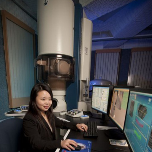
JEOL JEM-ARM200CF (Aberration Corrected Cold Field Emission Scanning Transmission Electron Microscope ) Heading link
Location: Science and Engineering South, 104A
Description: The JEM-ARM200CF is a probe aberration corrected 200kV STEM/TEM
with a cold field emission source with 0.35eV energy resolution. For
HAADF imaging at 200kV this instrument has a resolution of less than
0.08nm. It was installed in 2011.
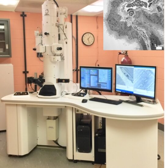
JEOL JEM 1400 Flash TEM Heading link
Location:
Description: The JEOL JEM 1400 Flash TEM is a versatile 120kV Transmission Electron Microscope (TEM) which offers a wide range of applications and can be easily optimized for users’ research purposes. It is known for its ease of use with a multi-touch screen to provide high resolution imaging and analysis for a variety of interests, such as biologicals, polymers, nanotechnology, quality control, and advanced materials development.
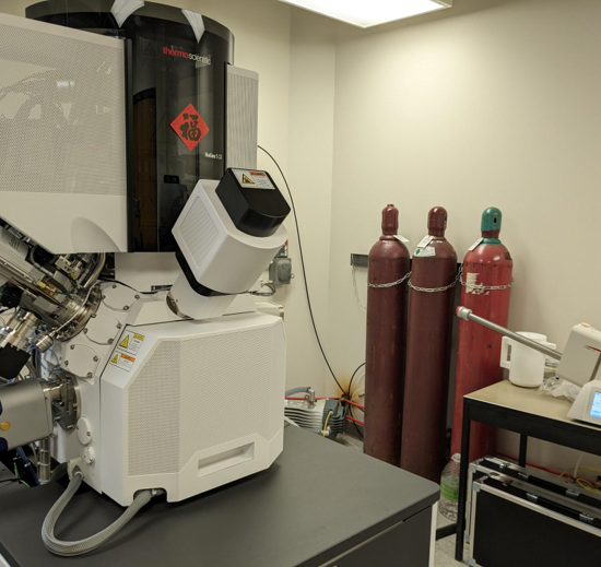
ThermoFisher Scientific Helios 5CX Focused Ion Beam (FIB) Heading link
The Thermo Scientific™ Helios™ 5 CX DualBeam is part of the fifth generation of the industry-leading Helios DualBeam family. It is carefully designed to meet the needs of scientists and engineers, combining the innovative Elstar™ electron column for ultra high-resolution imaging and the highest materials contrast with the superior Thermo Scientific™ Tomahawk HT Focused Ion Beam (FIB) Column for the fastest, easiest and most precise high-quality sample preparation. In addition to the most advanced electron and ion optics, the Helios 5 CX DualBeam incorporates a suite of state of-the-art technologies that enables simple and consistent high resolution S/TEM and Atom Probe Tomography (APT) sample preparation, as well as the highest-quality subsurface and 3D characterization, even on the most challenging samples.
- Auto TEM—automation of TEM lamella preparation
- Auto Slice and View– 3D construction with images, EDS and EBSD data
- Maps3.0– automated montages of imaging of large area of specimens (can be wafer scale). Ideal for biological specimens.
- W, Pt and C deposition sources
- Cryo transfer capability with Leica VCT/VCM transfer station– for air-sensitive materials such as Li and biological specimens
- TEM thin lamella specimen prep
- EDS and EBSD detectors—chemical composition & structural information
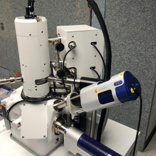
JEOL JSM-IT500HR (Field Emission Scanning Electron Microscope (FESEM)) Heading link
Location: Science and Engineering South (SES), 104C
Description: The IT500HR has a high-brightness electron gun system delivering high-resolution Field Emission performance, and is a Variable Pressure SEM. Its large analytical chamber with multiple ports accommodates a variety of detectors for imaging and analysis; secondary (SED) and backscatter (BSED) electron detectors, and energy dispersive spectroscopy (EDS) and electron backscatter diffraction (EBSD) techniques.
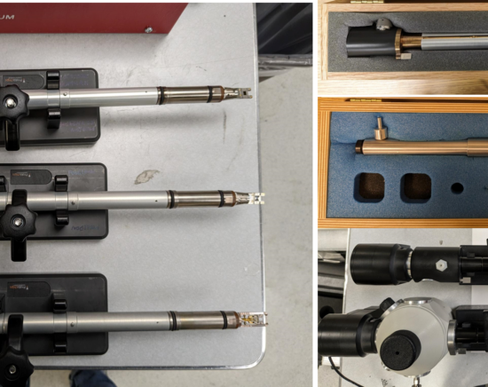
In-Situ Stages (Special Holders for Transmission Electron Microscopy) Heading link
Location: Science and Engineering South, 116A, 104B
Description: As well as the standard JEOL single and double tilt holders, EMC also has a number of special holders designed for the JEM-ARM200CF. There is no additional charge to use these holders in addition to the hourly microscope use charge, but regular users must book the holders on iLab. Some holders need conditioning the day before use and you will need additional training to use any of these holders – please e-mail EMC staff in advance if you want to use these holders.
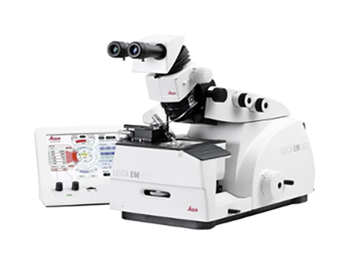
Leica UC7 Ultramicrotome Heading link
Prepare high-quality ultra- or semi-thin sections for your transmission electron or light microscope investigation whilst simultaneously creating perfectly smooth block face surfaces for atomic force, scanning electron, or incident light microscopy. For ultrathin cryo- sections or surfacing of cryogenic material, you can equip your EM UC7 ultramicrotome with the EM FC7 low-temperature sectioning system within minutes.
– Touch-sensitive-control unit
– Biological TEM thin section (<100 nm) and semi-thin section (<1 um) preparation
– Can be upgraded to Cryo ultramicrotome
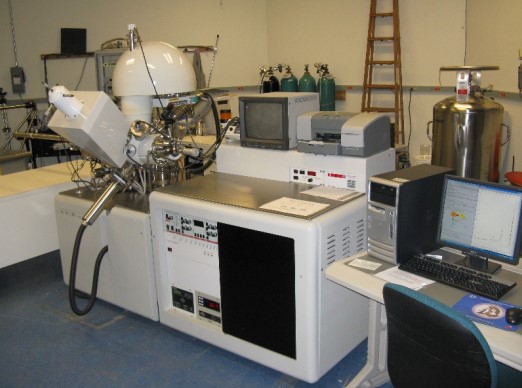
Kratos AXIS-165 (X-Ray Photon Spectroscopy) Heading link
Location: Science and Engineering South, 109B
Description: The Kratos AXIS-165 Surface Analysis System is a multi-technique instrument.
Our instrument, installed in December 2004, is equipped for X-ray Photon Spectroscopy (XPS – also known as ESCA (Electron Spectroscopy for Chemical Analysis)). This technique is surface sensitive (less than 8nm for XPS) with a spatial resolution in X and Y of down to 30 µm.
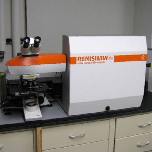
Renishaw inVia Reflex (Micro-Raman Spectrometer) Heading link
Location: Science and Engineering South, 116B
Description: The Renishaw inVia Reflex Raman, installed in June 2016, has green 532nm/50mW diode pumped solid state laser and red 633nm/17.5mW HeNe laser and has auto switch and alignment of the lasers. The Microscope is a research grade Leica DM2700M microscope with better than 2.5um depth resolution using a 100x objective with 0.35mm working distance for maximum confocal performance, a 50x objective with 0.5mm working distance, a 20x objective with 1.15mm working distance and a 5x objective with 14mm working distance.
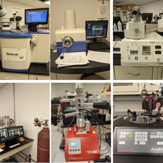
Conventional TEM and SEM specimen preparation instruments Heading link
Conventional TEM and SEM specimen preparation instruments:
Fischione 1050 Ion Mill
Fischione 1040 Nano Mill
JEOL SM-09010 Cross Sectional SEM polisher
South Bay Tech Plasma Cleaner with Ar and O2
Dry Pumping stations for storage of holders
Allied High Tech polishing wheel for TEM specimens